
Colour in gemstones is mainly due to the selective absorption of wavelengths by the transition elements present in their structure.
- In order to discover which wavelengths have been absorbed by a gemstone, it is necessary to inspect it using an instrument called the spectroscope.
- When light is transmitted through or reflected from a gem material, some colours of light representing certain wavelengths may be selectively absorbed.
- Any wavelengths that are selectively absorbed will appear as dark vertical lines, bands or cutoffs of varying width and intensity across the range of spectral colours known as the absorption spectrum.
- An emission spectrum is observed when the elements emit light instead of absorbing the incident radiation which is seen as bright vertical fluorescent lines.
Spectroscope (Visible Range): Gemmological spectroscopes fall into two categories, prism spectroscope and diffraction grating spectroscope. The basic difference between the two lies in the brightness of the spectrum and the absorption spectrum.
- Diffraction Grating Spectroscope: Wavelengths are evenly distributed, multiple spectral images – bright
- Prism Spectroscope: Red end is compressed while the blue end is expanded, purer spectrum.
Commonly a prism spectroscope is used.
- This instrument consists essentially of a train of prisms enclosed in a tube, with a slit at one end and a convex lens at the other.
- A converging lens is placed between the slit and the prism assembly to collimate (make parallel) the light.
- The spectroscope separates white light into its component spectral colours, which range in wavelength from 400nm to 700nm.
- These absorption patterns are characteristic to varying degrees, of some gem materials and can provide valuable information in their identification.
Uses of spectroscope
- Identification of gemstones by absorption spectrum.
- To detect dye treatments.
- Get an indication of colouring elements present in the gemstone.
Procedure:
- Place the stone on the lighted base. (Only light that passes through the stone should enter the slit of the spectroscope).
- Position and focus the light source so that the maximum amount of light is transmitted through the stone (for transparent stones) or reflected (for opaque stones) from the surface.
- Close the slit width of the spectroscope completely; then open it slowly until the entire spectrum is visible. Observe and note the cutoff, bands and lines at various positions.
- Examine the stone in different directions, as some stones exhibit directional absorption spectrums.
- Make a conclusion by considering all the lines and bands seen in each different direction.
- The nature of the spectrum observed is affected by the lighting, transparency of the stone, depth of colour etc.
- Dust or dirt cause dark horizontal lines, across the spectrum.
Transition Elements
| Element | Colour | Spectrum |
|---|---|---|
| Vanadium | Usually plays secondary role. Green: beryl, tsavorite, color changing sapphire, tanzanite. | Weak absorption in the blue and green portions. |
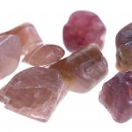
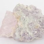
| Chromium | Red: ruby, spinel, pyrope garnet. Green: emerald, demantoid garnet, jadeite, alexandrite. | Broad absorption in violet and green with narrow lines in the red area. |
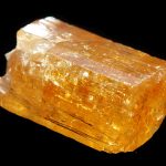
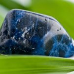
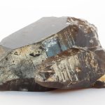
| Iron | Yellow: chrysoberyl, citrine. Green: sapphire, peridot, tourmaline. Blue: aquamarine, spinel, sapphire. Red: almandine garnet. | Broad absorption bands in blue-green portions of the spectrum. |
| Nickel | Green: chrysoprase, prase opal. | Normally no absorption pattern, when present, seen. |
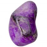
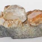
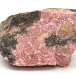
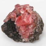
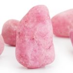
| Manganese | Pink: rhodochrosite, rhodonite, morganite, kunzite. | Broad absorption bands in the violet and blue portions. |
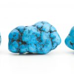
| Copper | Blue: chrysocolla, azurite, turquoise. Green: malachite, dioptase. | Very faint absorption if any, in the blue area. |
| Cobalt | Blue: synthetic spinel, glass, synthetic blue quartz. | Three bands in the green, yellow and orange portions. |
| Titanium | Occasionally occurs with Iron. Blue: sapphire, benitoite. | Absorption is with iron in the blue. |
| Rare Earth (Neodymium, Praesodymium) |
Yellow: apatite, danburite. Green: sphene. | Several sharp bands in the yellow region. |
The Light Spectrum
| Color | Angstrom Units (A.U.) | Nanometers (nm) |
|---|---|---|
| Ultra-Violet | 1000 – 3900 | 100 – 390 |
| Violet | 3900 – 4300 | 390 – 430 |
| Blue | 4300 – 4900 | 430 – 490 |
| Blue-Green | 4900 – 5100 | 490 – 510 |
| Green | 5100 – 5500 | 510 – 550 |
| Yellow-Green | 5500 – 5750 | 550 – 575 |
| Yellow | 5750 – 5900 | 575 – 590 |
| Orange | 5900 – 6300 | 590 – 630 |
| Orange-Red | 6300 – 6500 | 630 – 650 |
| Red | 6500 – 7000 | 650 – 700 |
| Deep Red | 7000 – 7800 | 700 – 780 |
| Infra Red | 7800 – 10,000,000 | 780 – 1,000,000 |


Leave a Reply
You must be logged in to post a comment.