
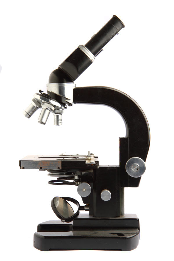
This consists mainly of a mechanical section and an optical section. Following is a brief description of some of the main parts and their functions.
Mechanical section:
- The base or foot has to be fairly solid to maintain the overall balance.
- The vertical limb contains the focal adjustments; knob for moving the body tube and at the lower end is the stage on which the stone is placed.
- The stage has a central aperture through which light from below can pass, a diaphragm to control the light aperture, holders to place the tweezer and a knob to provide the required illumination (dark or light field).
Optical section:
- The body tube contains the optical components.
- At the bottom end of the tube is the objective which is made of compound lenses to provide the primary magnified image.
- At the top there are two oculars (eyepieces) with a specific magnification to produce an enlarged virtual image of the image formed by the objective.
- A combination of lenses produces the enlarged image (zoom facility).
There are a few important features of a microscope which must be considered:
- The field of view. The greater the magnification of the microscope, the smaller will be the field of view and the working distance.
- The working distance.
- The depth of focus i.e. the distance over which the image is within focus becomes smaller as the magnification increases.
- Adjust the microscope eyepieces (oculars) to give you a single three dimensional view.
- Clean the stone thoroughly with a lint-free stone cloth.
- Hold the stone with the stone holder and fix it to the stand provided.
- Using low magnification, observe the stone in all directions and adjust the focus till the stone is visible clearly. Look for fractures, structure, lustre, inclusions etc. by focusing up or down.
- Switch to higher magnification and attempt to identify the inclusions, cleavage etc.
- Observe the distinct differences in surface features and internal features.
- Proper illumination of the stone helps in correct identification.
Immersionscope
Observing a stone by immersion is useful since it eliminates surface and facet reflections and enables one to observe the internal features clearly.
- The immersionscope consists mainly of a stage on which the microscope, transparent immersion cell and light source with iris diaphragm are placed in a horizontal plane.
- Choose the appropriate liquid for immersion. In general, liquids having a similar refractive index as the stone are used. E.g. benzyl benzoate (R.I. 1.54) is used for emeralds and quartz, water (R.I. 1.33) for opal, while methylene iodide (R.I. 1.74) is used for Rubies and Sapphires.
- Solid and liquid inclusions – like crystals, nature of fingerprints and phase inclusions and their optic character can be examined more clearly.
- Growth features like plato lines, Brazil law twinning, graining, hound’s tooth markings, growth zoning, curved lines / bands etc. can be easily observed.
- Type of enhancement – diffusion treatment, fracture filling etc. In diffusion, blue colour concentrations on facet edges and colour bleeding. Fracture filling with colourless oils / resins exhibits typical colour flashes on rotation and coloured fillings exhibit colour concentrations in fractures.
- Composite stones show a distinct junction plane and the inclusions in the two portions.
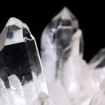
- Commonly gemstones like emerald, opal, quartz, ruby and sapphire are examined with an immersionscope.
Precautions:
- Appropriate liquid should be used for better clarity.
- Before doing the immersion test, confirm that the stone is not porous.
- Dyed or coated since it may absorb the solution and thus be decolourised / damaged.
- In the case of composites, care should be taken to prevent the joined parts from separating.
- Do not inhale the liquid.
- Clean the stone immediately after the immersion test with absorbent cloth / paper.
Magnification: Magnifying an object simply means to observe it as a larger image, which helps in observing the finer points. Magnification is important in that it is useful in various aspects of gemstone identification.
- External features:
- Identification with respect to rough and cut gemstones.
- Identification of surface features like markings on rough crystals, polishing lines, pits, nicks etc. on cut stones.
- Identification of habit and form in rough and the type and quality of cut in facetted stones.
- Identification of surface enhancements like coating, foiling, diffusion treatment etc.
- Identification of phenomenon.
- Internal features:
- Identification of inclusions which would be useful in determining.
- Natural and synthetic stones.
- Geographic Origin of stones.
- Identification of a species.
- Detection of composites.
- Grading of gemstones: This is internationally standardised for diamonds using 10x magnification as the standard, while coloured stones are graded more on individual office requirements.
- Jewellery: Observation of quality of jewellery in terms of setting, texture etc.
Illumination and magnification: This is of great importance since it determines the visibility of the features to a great extent.
- Most microscopes are provided with a built-in means of illuminating the specimen on the stage. This can be as basic as a lamp and condenser lens assembly under the stage, with an iris control to vary the area of illumination.
- A model of a 10X lens has been made with an attached dark field illumination.
- In the more sophisticated models, the choice of incident, light-field, or dark-field illumination is provided, the latter two being contained in the sub stage lamp assembly.
- Light-field illumination: Light is transmitted upwards through the specimen and into the objective of the microscope. An inclusion appears as a dark object against a bright background. This is useful while viewing the internal features in opal.
- Dark-field illumination: Light is directed into the gem from the sides, and there is no direct light path between the lamp and the objective. Dark-field illumination is generally the preferred method for gemmological work, as it gives better contrast. An inclusion appears as a bright object against a dark background.
- Diffused illumination: It is produced using a diffuser type tissue paper or translucent plaster etc. Curved coloured bands, curved lines, and colour zoning are seen clearly even in light coloured stones.
- Polarised illumination: It is used to detect the optic character of gemstones, or of inclusions in a gemstone, to detect growth features like plato lines and Brazil law twinning.


Leave a Reply
You must be logged in to post a comment.